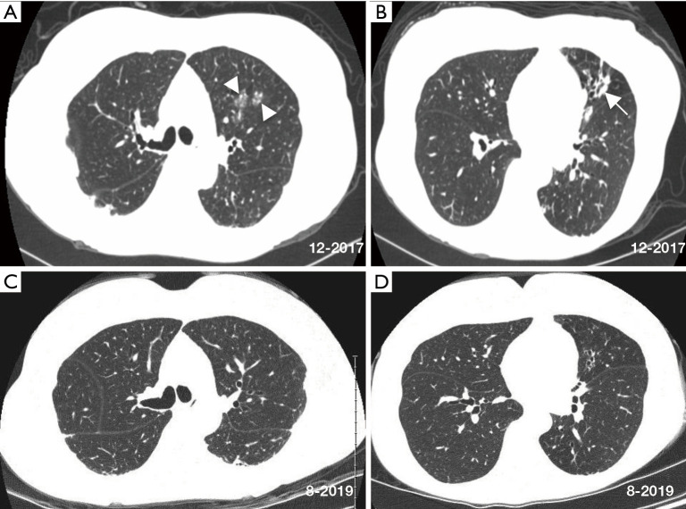
Figure 1.
Axial chest CT scan images at the time of referral show relatively mild centrilobular nodules/ground glass opacities (arrowheads) and bronchiectasis (arrow) affecting the left lung more than the right lung (A,B). Chest CT scan nearly 2 years later (C,D) show modest radiographic improvement with resolution of the left upper lobe ground glass opacity, decrease in subpleural densities, and minimal to modest improvement of the lingula following antibiotic treatment for NTM, use of airway clearance measures, implementation of head-of-bed elevation and other anti-reflux measures, and reduction of soil aerosol exposures. CT, computed tomography; NTM, Non-tuberculous mycobacterial.

