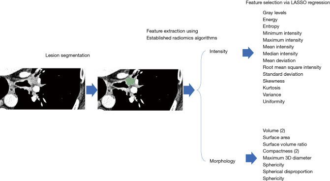
Figure 2.
Radiomics workflow. The cancer is first segmented in 3Ds, allowing distinction from other adjacent structures. This 3D structure is then analyzed using established radiomics algorithms related to either Morphology or Intensity. These radiomics features are then evaluated via LASSO regression to determine those most highly associated with response to the therapy. 3D, 3-dimension; LASSO, least absolute shrinkage and selection operator.

