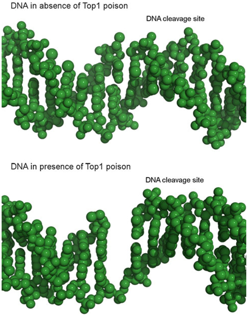
Fig. 1.

Comparison of DNA cleavage sites in the absence and presence of a Top1 poison. DNA is shown as green spheres. DNA structures obtained from crystal structures of covalent Top1-DNA complexes: PDB 1K4S (top panel, no ligand), and 1K4T (bottom panel, ligand bound) [27]
