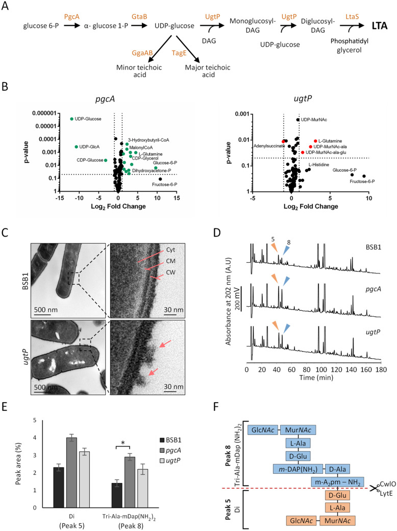
Figure 1.
The ugtP mutant has altered cell surface and PG composition. (A) Teichoic acid glycolipids pathway of the cell wall in B. subtilis. DAG, diacylglycerol. (B) Volcano plot for data generated by metabolomics analysis for BSB1 ΔugtP and BSB1 ΔpgcA showing increased levels of several lipid II precursors (C) Transmission electron microscopy (TEM) images showing a rougher cell surface structure in cells lacking UgtP compared to wild type cells. Cyt cytoplasm, CM cytoplasmic membrane, CW cell wall. Arrows indicate the rough cell surface. (D) HPLC analysis of muropeptides isolated from BSB1 ΔugtP and BSB1 ΔpgcA mutants showed increased levels of Di (muropeptide 5) and Tri-Ala-mDap(NH2)2 (muropeptide 8) muropeptides compared to BSB1 suggesting increased endopeptidase activity. (E) Diagram representing the relative quantification of the two muropeptides in BSB1, BSB1 ΔugtP and BSB1 ΔpgcA. *p ≤ 0.05. (F) The scheme represents the structure of the two muropeptides and the cleavage site of the LytE and CwlO endopeptidases.

