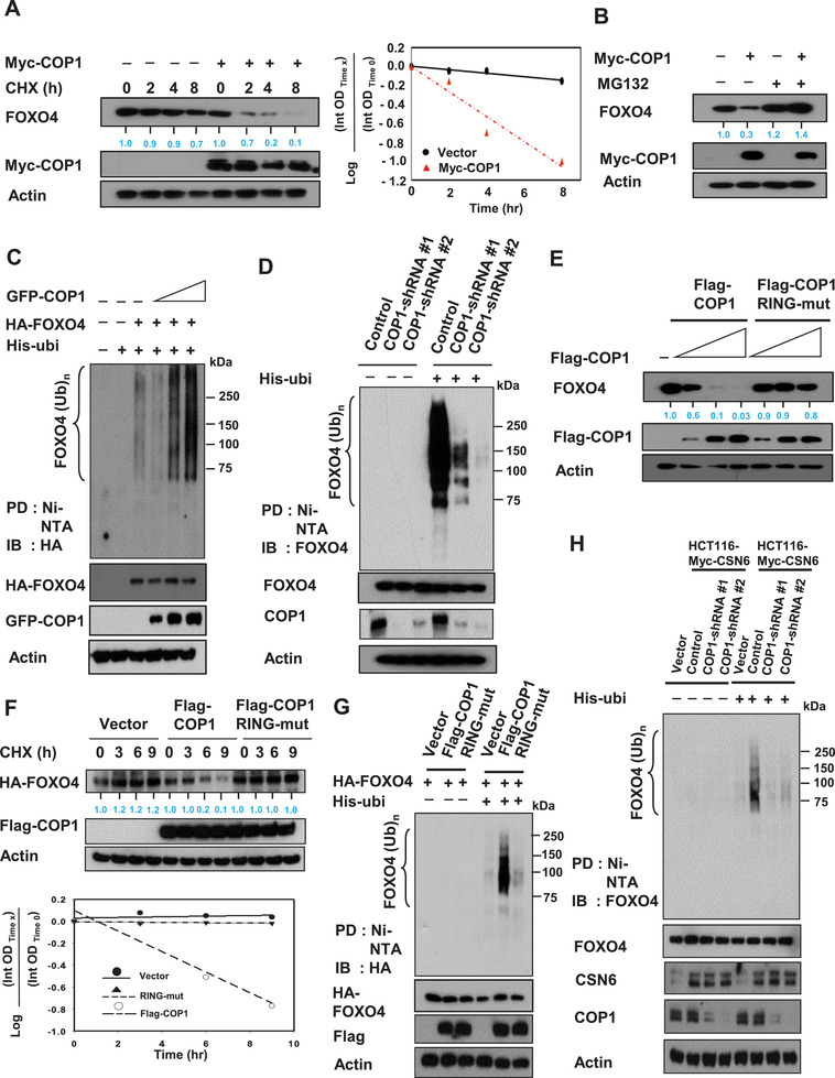
Figure 3.

COP1 is involved in CSN6‐mediated ubiquitination of FOXO4. A) SW480 cells transfected with the indicated expression vectors were treated with cycloheximide (CHX) (100 µg mL−1). Cell lysates were immunoblotted with indicated antibodies. The turnover rate of FOXO4 is shown. B) SW480 cells transfected with either Myc‐COP1 or vector control was treated with proteasome inhibitor MG132. Lysates were immunoblotted with indicated antibodies. C,D) 293T cells transfected with C) the indicated plasmids or HCT116 cells infected with D) COP1 shRNA were treated with 5 µg mL−1 MG132 (Sigma) for 6 h before harvesting. Cells were lysed in guanidine–HCl containing buffer. The cell lysates were then pull down (PD) with nickel beads Ni‐NTA and immunoblotted with the indicated antibodies. E) DLD1 cells were transfected with the indicated expression vectors. Lysates were immunoblotted with indicated antibodies. F) 293T cells were transfected with the indicated expression vectors. The cells were treated with cycloheximide (CHX) (100 µg mL−1) for the indicated hours. Cell lysates were immunoblotted with indicated antibodies. The turnover rate of FOXO4 is shown. G) 293T cells were transfected with the indicated expression vectors. Cells were treated with MG132 for 6 h before harvesting, and lysed in denaturing buffer (6 m guanidine‐HCl). The cell lysates were then pull down (PD) with nickel beads and immunoblotted with anti‐HA. H) Myc‐CSN6 overexpressing HCT116 cells infected with either COP1 shRNA were treated with MG132 for 6 h before harvesting. HCT116 vector cells were used as a control. Cells were lysed in denaturing buffer (6 m guanidine‐HCl). The cell lysates were then pull down (PD) with nickel beads and immunoblotted with indicated antibodies.
