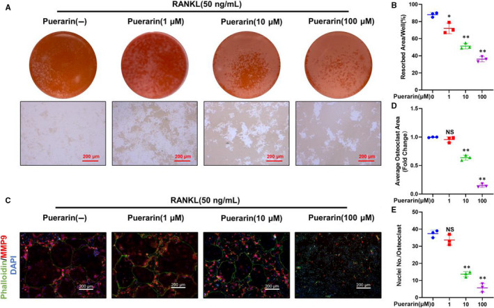
Figure 4.

Puerarin influenced osteoclast bone resorption and F‐actin ring formation in vitro. (A) Representative images of bone resorption. (B) Quantification of resorption area/well. (C) Representative images of cells stained with phalloidin, MMP9 and DAPI. (D and E) Quantification of the average osteoclast area and number of nuclei/osteoclast. n = 3; scale bar = 200 μm; NS: Not statistically significant, *P < .05, **P < .01, * vs the 0 μmol/L group
