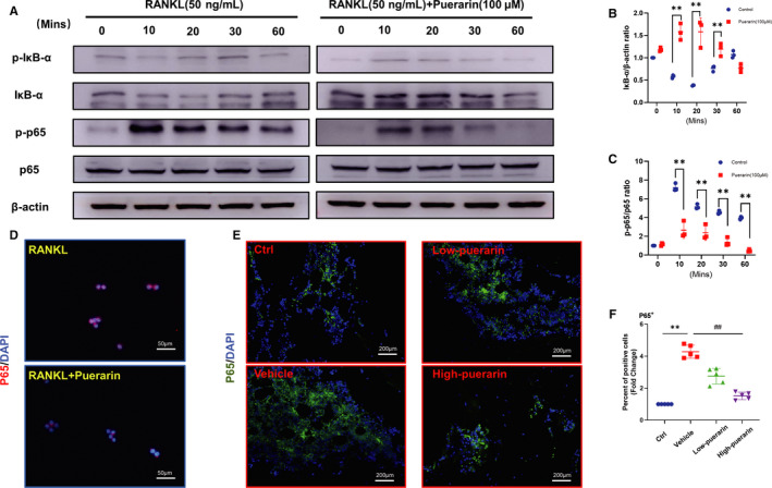
Figure 6.

Puerarin suppressed the activation of the NF‐κB signalling pathway during osteoclastogenesis. (A) Cell lysate was subjected to Western blotting with antibodies against phosphor‐IκB‐α, IκB‐α, phosphor‐p65 and p65. (B and C) The ratio of IκB‐α/β‐actin and p‐p65/p65, n = 3, **P < .01, * vs control group. (D) Representative images of p65 in the nucleus, scale bar = 20 μm. (E) Representative images of immunofluorescence staining for p65. (F) Quantification of p65‐positive cells. n = 5; **P < .01, ##P < .01, * vs the Ctrl group, # vs the vehicle group
