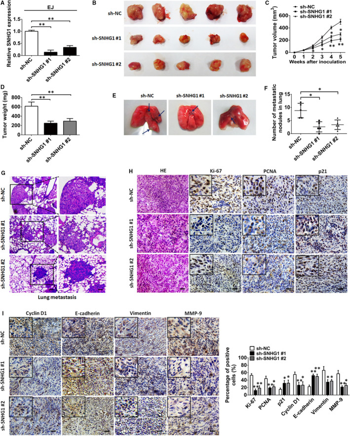
FIGURE 3.

SNHG1 silencing suppresses tumour growth and metastasis in vivo. A, SNHG1 expression levels were assessed by RT‐qPCR in EJ cells harbouring a stable transfection with sh‐SNHG1 #1 or sh‐SNHG1 #2 vectors. B, Representative images of tumour xenografts in nude mice subcutaneously injected with sh‐SNHG1 #1‐ or sh‐SNHG1 #2‐transfected EJ cells after 4 weeks. Tumour volume (C) and tumour weight (D) were compared between the sh‐NC and sh‐SNHG1 groups. E, Representative sections of pulmonary metastatic mouse models are shown. The blue arrows indicate the tumour nodules. F, The number of metastases in the lung was assessed. G, Representative pathological images of the metastatic nodules stained by HE in the lungs. Scale bar indicates 200 μm in upper layer; 50 μm in lower layer. H‐I, Representative images of HE and immunochemistry staining for Ki‐67, p21, PCNA, cyclin D1, E‐cadherin, vimentin, and MMP‐9 in xenograft tumour tissues of mice in the SNHG1‐silenced group (sh‐SNHG1 #1 or sh‐SNHG1 #2) and the control group (sh‐NC). Percentage of positive marker expression of analysed marker genes was also assessed. Scale bar, 50 μm. * P < .05 and ** P < .01
