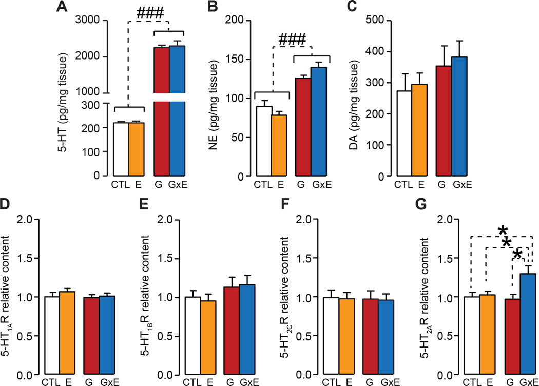
Figure 8. G×E pups show elevated 5-HT2A receptor staining in the orbitofrontal cortex at postnatal day 7.
(A-B) Digital light micrographs and percent intensity of staining of 5-HT2A receptor immunopositive cells in the orbitofrontal cortex (C-D) Digital light micrographs and percent intensity of staining of 5-HT2A receptor immunopositive cells in the temporal-occipital cortex at postnatal day 8. Immunohistochemical studies revealed that (A-B) 5-HT2A levels were significantly enhanced in the orbitofrontal cortex of stress-subjected MAOANeo pups (3-way ANOVA; n=7/group); conversely (C-D), no differences were found in the temporal-occipital cortex (3-way ANOVA; n= 7/group). Data are shown as means ± SEM. Scale bar is set at 25 μm. *, P<0.05 for all comparisons indicated by dotted lines (interactions). Abbreviations: CTL, unstressed wild type (WT) mice; E, WT mice subjected to early stress during the first postnatal week; G, unstressed MAOANeo mice; G×E, MAOANeo mice subjected to early stress during the first postnatal week.

