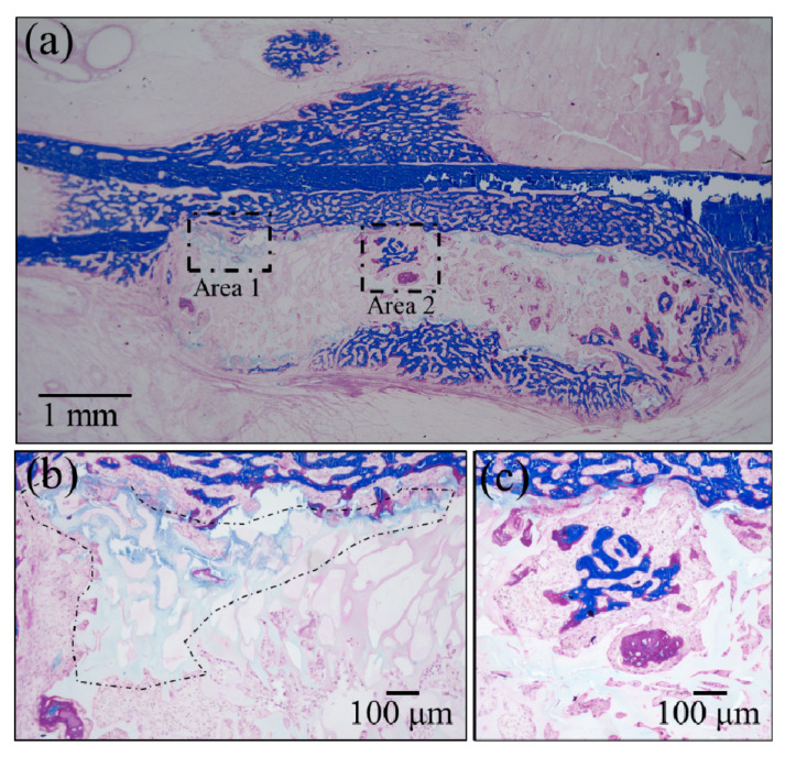
Figure 6.
Results of Masson’s trichrome staining: (a) low-resolution histological image of the regenerated bone treated with the NanoCliP-FD scaffold; enlargements of the square insets are shown in (b) and (c) for Area 1 and Area 2, respectively; mature bone in deep blue, collagen in light blue, and immature bone in violet colors.

