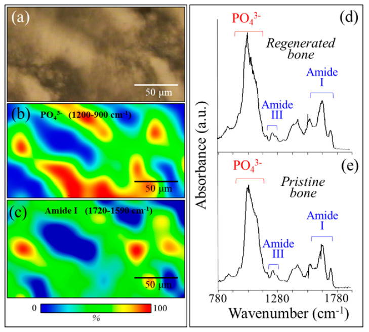
Figure 8.
(a) Bright-field optical micrograph and related synchrotron radiation–based Fourier transform infrared (SR-FTIR) image of (b) apatite and (c) collagen structures in a microscopic portion within the regenerated bone scaffold area. In (d) and (e), the SR-FTIR spectra of regenerated bone are shown in correspondence with the scaffold and pristine cortical bone, respectively.

