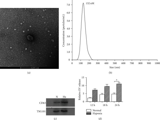
Figure 1.

Characterization and quantification of exosomes produced by KEL cells under hypoxia or normoxic condition. KEL cells were incubated under hypoxic or normoxic condition for 12 h, 18 h, or 24 h to collect exosomes. (a) Representative electron micrograph of exosomes isolated from hypoxia- or normoxia-conditioned medium of KEL cells. Scale bar, 200 nm. (b) A representative plot showing the size distribution of exosomes was carried out using nanoparticle tracking analysis (NAT). (c) Western blot analysis showing the presence of CD63 and TSG101 in exosomes derived from KEL cells. (d) Quantification of exosomes produced by KEL cells under hypoxia or normoxic condition at various time points (12 h, 18 h, and 24 h). The measurements were done using the NTA system in triplicate, and data presented as fold change of the EV number. N: normoxic; Hx: hypoxic. ∗P < 0.05.
