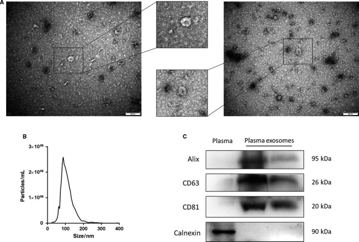
FIGURE 2.

Characterization of plasma exosomes. A, Representative electron micrograph of plasma exosomes. B, Size distribution of plasma exosomes analysed by nanoparticle tracking system. C, Western blot analysis of common exosomal markers Alix, CD63 and CD81, and the endoplasmic reticulum marker calnexin. Plasma was used as a control.
