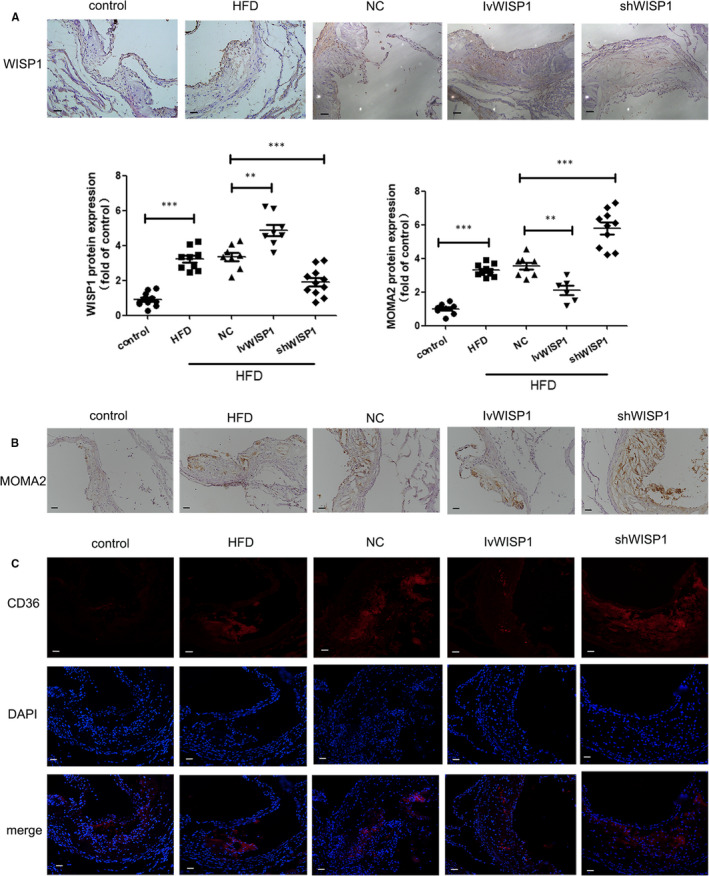
Figure 3.

WISP1 leads to lipid deposition and recruitment of macrophages in ApoE‐/‐ mice. Immunohistochemical staining of WISP1 (A) and MOMA2 (B) from the aortic sinus (scale bar: 50 μm). (C) Immunofluorescent staining of CD36 in the aortic sinus (scale bar: 50 μm) (*P < .05, **P < .01, ***P < .001; data = means ±SD). HFD, high‐fat diet; NC, null lentivirus; IvWISP1, lentivirus WISP1; shWISP1, WISP1‐shRNA; WISP1, WNT1‐inducible signalling pathway protein 1
