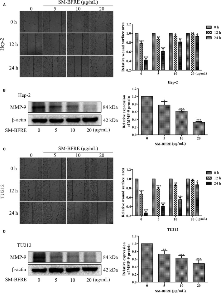
FIGURE 3.

Effect of SM‐BFRE on the migration of laryngeal carcinoma cells. (A and C) Micrographs of two laryngeal carcinoma cells at 0, 12 and 24 h after treatment with SM‐BFRE (0, 5, 10 and 20 μg/mL), respectively, and relative wound surface area in wound healing assay was calculated by ImageJ software. (B and D) The expression of MMP‐9 protein was detected by Western blot assay, and the relative expression was calculated by Image Lab software. β‐actin was used as a control. *P < .05, **P < .01 or ***P < .001 compare to control (0 μg/mL served as control)
