
FIGURE 5.
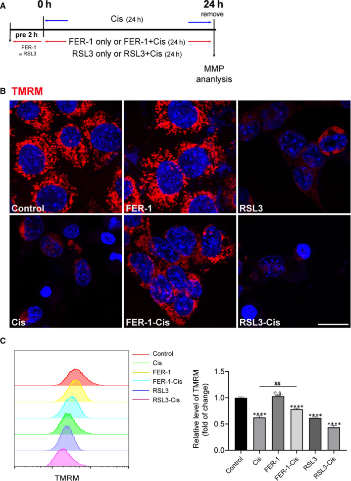
Effect of ferrostatin‐1 (FER‐1) on mitochondrial membrane potential (MMP) in cisplatin‐damaged House Ear Institute‐Organ of Corti 1 (HEI‐OC1) cells. A, The experimental workflow. The HEI‐OC1 cells were pre‐treated with 3 μmol/L RSL3 or 30 μmol/L FER‐1 for 2 h and then treated with or without 30 μmol/L cisplatin for another 24 h, or treated with 30 μmol/L cisplatin alone for 24 h, and then MMP was detected by TMRM. B, Representative images of HEI‐OC1 cells stained with TMRM. Scale bar, 20 µm. C, Measurement and quantification of MMP by flow cytometry. The data are shown as mean ± SEM. of three independent experiments. ****P < 0.0001 and n.s. no significant vs the control group; ## P < 0.01 vs the cisplatin group