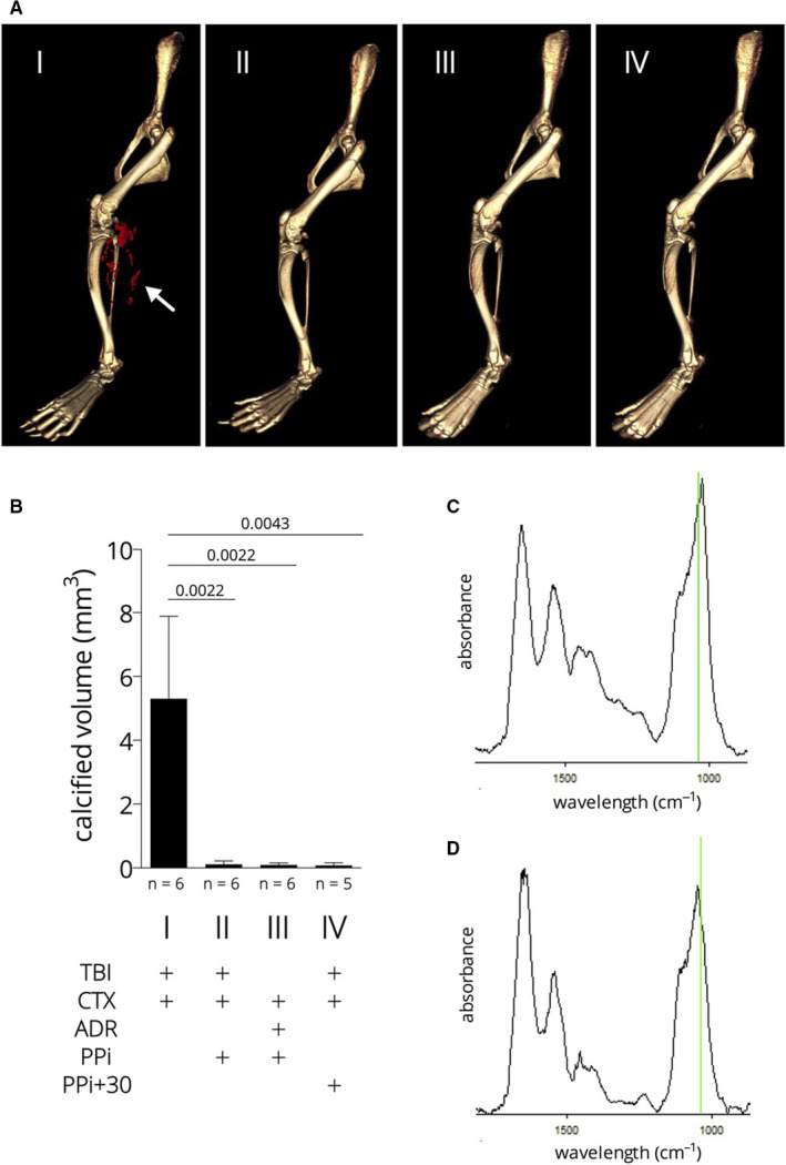
FIGURE 4.

Calcification of the hamstring muscle upon complex injury; effect of pyrophosphate. Panel A: micro‐CT images, white arrow points to the mineral deposits (red). Panel B: quantitative determination of the mineral deposits by micro‐CT volume measurement. Panel C and D: representative Fourier transform infrared microspectroscopy (μ‐FTIR) spectra of microcalcifications of the hamstring muscle of a TBI + CTX animal (C) and a TBI + CTX animal treated with pyrophosphate (D). Green lines correspond to 1030 cm−1, the ν3 P–O stretching vibration mode of apatite
