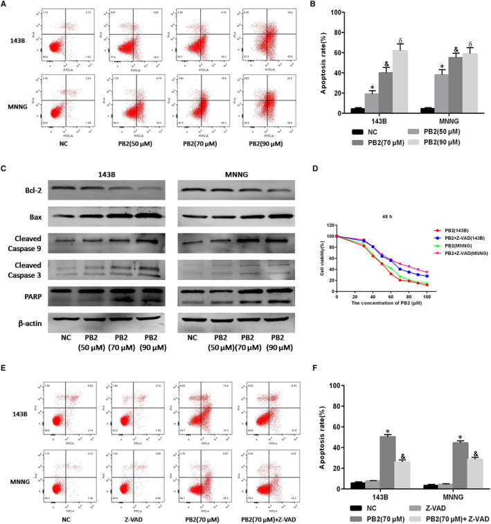
FIGURE 2.

Effects of PB2 on 143B and MNNG cell apoptosis. A,B, 143B and MNNG cells were treated with different concentrations of PB2 (0, 50, 70 and 90 μmol/L) for 48 h. Apoptosis of 143B and MNNG cells was determined by flow cytometry (n = 3, *,&,δ P < .001 for PB2 vs NC). C, Protein levels for Bcl‐2, Bax, cleaved Caspase‐9, cleaved Caspase‐3 and PARP assessed by Western blotting. D, 143B and MNNG cells were treated with PB2 or PB2 + Z‐VAD (50 μmol/L) for 48 h, and cell proliferation was determined by CCK‐8 assays. E,F, Apoptosis of 143B and MNNG cells determined by flow cytometry (n = 3, * P < .001 for PB2 vs NC; & P < 0.001 for PB2 vs PB2 + Z‐VAD)
