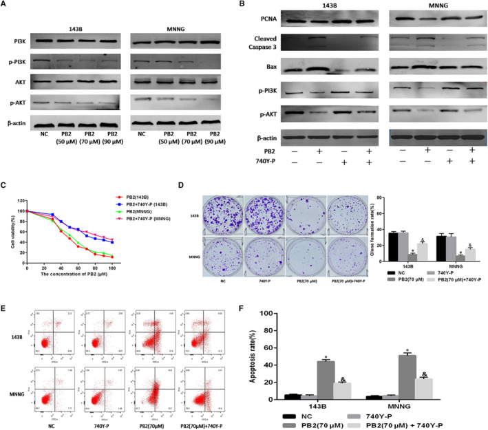
FIGURE 4.

The role of the PI3K/AKT pathway in OS cell proliferation and apoptosis. A, Expressions of PI3K, p‐PI3K, AKT and p‐AKT in 143B and MNNG cells determined by Western blotting. B, 143B and MNNG cells treated with PB2 or PB2 + 740Y‐P (20 μmol/L) for 48 h, and expression of PCNA, cleaved Caspase‐3, Bax, p‐PI3K and p‐AKT determined by Western blotting. C, Cell proliferation determined using the CCK‐8 assays. D, Macrograph of colony formation in 143B and MNNG cells (n = 3, * P < .05 for PB2 vs NC; & P < .05 for PB2 vs PB2 + 740Y‐P). E,F, Apoptosis of 143B and MNNG cells determined by flow cytometry (n = 3, * P < .05 for PB2 vs NC; & P < .05 for PB2 vs PB2 + 740Y‐P)
