FIGURE 3.

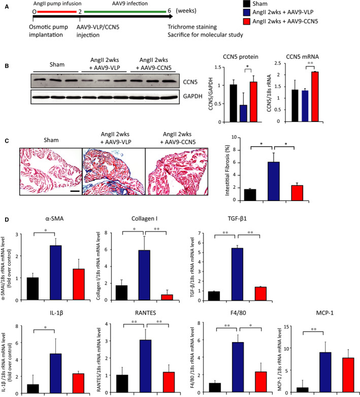
CCN5 reverses AngII‐induced atrial fibrosis in AAV9‐mediated CCN5 overexpressed mice. A, Experimental scheme is shown for B, C and D. Mice were underwent AngII‐infused osmotic pump implantation for 2 weeks. AAV9‐VLP or AAV9‐CCN5 (1 × 10¹¹ viral genomes per mouse) was injected via tail vein. 4 weeks later, hearts were analysed. (B) Total cell lysates from heart atrium were immunoblotted with antibodies against CCN5 and GAPDH (Left). Quantified protein level (Middle) and mRNA level (Right) of CCN5 are plotted. Heart was sectioned and stained with Masson's trichrome. Representative images are shown from left atrium. The percentage of interstitial fibrotic areas is plotted. Original magnification: 20 X, Scale bar: 200 μm (D) mRNA was isolated from left atrial and subjected to qRT‐PCR. Relative expression of pro‐fibrotic and pro‐inflammatory genes is shown. (n = 5 per group). Error bar =SD *P < 0.05, **P < 0.01
