Fig. 3.

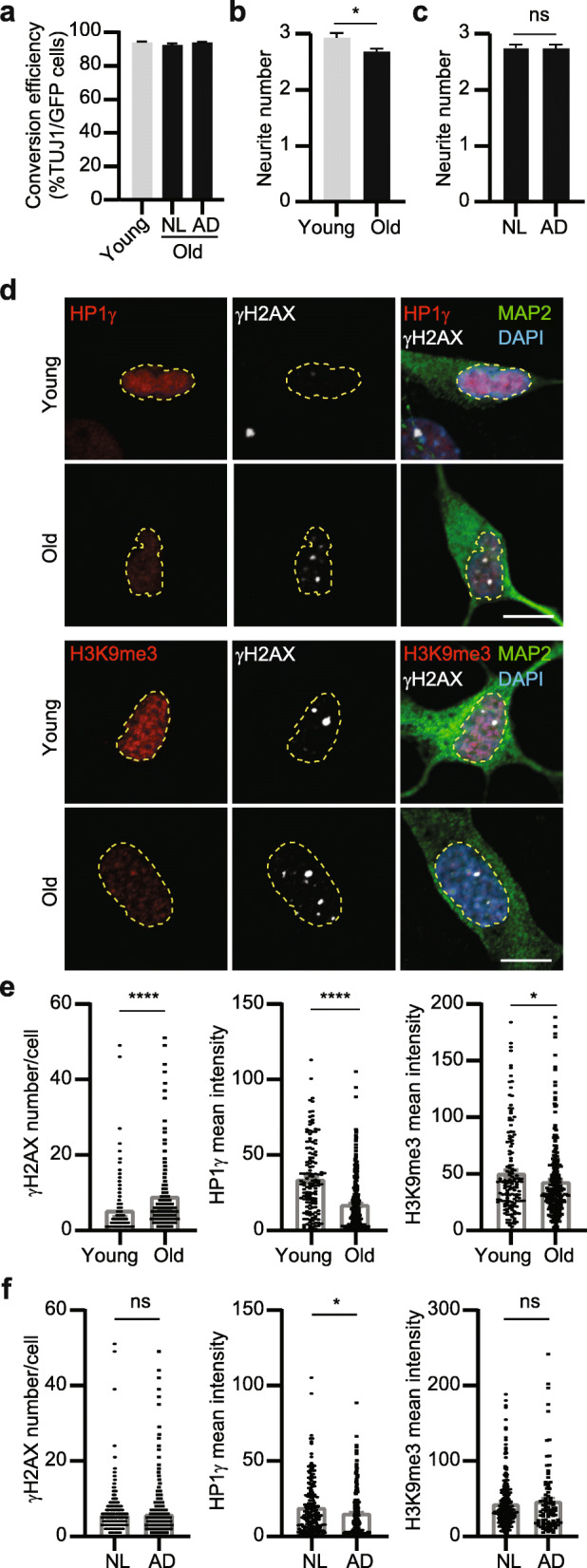
hiBFCNs retain aging-associated features. a Conversion efficiency for the indicated human fibroblast samples analyzed at 14 dpi. hiBFCNs were derived from young (2–3 years) and old (47–79 years) samples and co-cultured with mouse astrocytes (mean ± SEM; n = 3717 GFP+ cells for Young samples; n = 10,323 GFP+ cells for NL samples; and n = 8752 GFP+ cells for AD samples). b Quantification of primary neurite numbers per neuron for the indicated samples at 51 dpi (mean ± SEM; n = 361 cells for Young and n = 1052 for Old samples; *p = 0.0175). c Primary neurite numbers per neuron for the indicated samples at 51 dpi (mean ± SEM; n = 875 for NL and n = 538 for AD samples; ns, not significant). d Representative confocal images for marker expression in the indicated samples co-cultured with astrocytes at 51 dpi. The profiles of DAPI+ nuclei are outlined. Scale bar, 10 μm. e Quantifications of marker expression for the indicated young or old samples. Each dot represents a single cell (mean ± SEM; γH2AX: n = 426 for Young, n = 1263 for Old, ****p < 0.0001; HP1γ: n = 139 for Young, n = 421 for Old, ****p < 0.0001; H3K9me3: n = 146 for Young, n = 243 for Old; *p = 0.0446). f Quantifications of marker expression for the indicated NL or AD samples. Each dot represents a single cell (mean ± SEM; γH2AX: n = 621 for NL, n = 642 for AD; HP1γ: n = 200 for NL, n = 221 for AD, *p = 0.0162; H3K9me3: n = 243 for NL, n = 93 for AD; ns, not significant)
