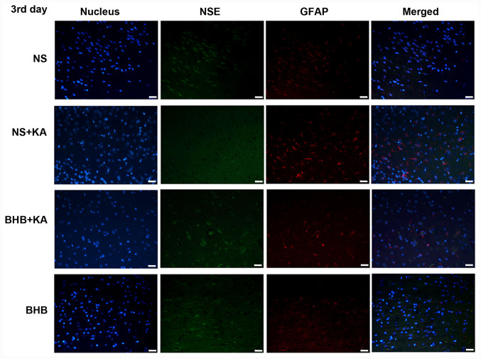
Figure 2.
Immunofluorescence staining of NSE and GFAP in the hippocampus tissues of rats 3 days after KA injection. Green signals represent NSE, red signals represent GFAP and blue signals represent the cell nuclei stained with DAPI. Scale bar, 50 µm. NSE, neuron specific enolase; GFAP, glial fibrillary acidic protein; BHB, β-hydroxybutyrate; KA, kainic acid; NS, normal saline.

