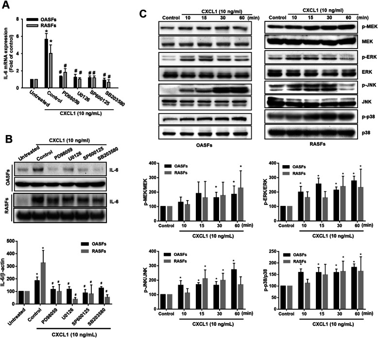
Fig. 4.
MAPK activation is required for IL-6 expression in response to CXCL1 in OASFs and RASFs. a, b OASFs and RASFs were pretreated with different inhibitors that block activation of various MAPK signal components (ERK, PD98059, 5 μM; MEK, U0126, 3 μM; JNK, SP600125, 3 μM; p38, SB203580, 5 μM) for 1 h, then incubated with CXCL1 (10 ng/mL) for 24 h. IL-6 expression was examined by qPCR and Western blot. The quantification of Western blot is provided in the lower panel. c The cell lysates were collected from OASFs and RASFs as described in Fig. 3c, and Western blot analysis assessed MEK, ERK, JNK, and p38 activation by monitoring the phosphorylated forms of these proteins. The quantification of Western blot is provided in the lower panel. (In the above experiments, OASFs; n = 10, RASFs; n = 10). Results are expressed as the mean ± SEM. Statistical analysis was conducted by using one-way ANOVA followed by Fisher’s LSD post hoc comparisons tests. *p < 0.05 compared with the respective groups in all figures (control and untreated); #p < 0.05 compared to the groups with control pretreatment followed by CXCL1 incubation

