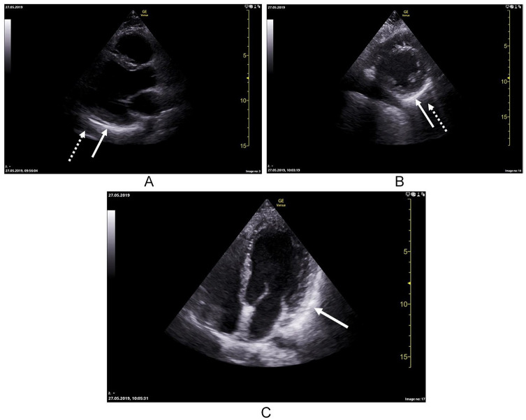
Figure 3.
Parasternal long axis demonstrating highly echogenic pericardium covering the inferolateral wall of the left ventricle (whole arrow) and a hypoechogenic area in front of it (dotted arrow) (A). Parasternal short axis showing the same features as long axis (B). Apical 4-chamber view showing highly echogenic pericardium covering the lateral wall of left ventricle (C).

