Figure 4.
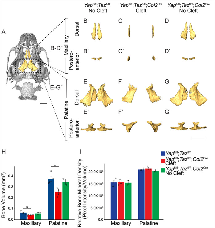
Deletion of Yap/Taz in the palatal shelf mesenchyme results in decreased volume of the bones of the secondary palate. (A) Ventral view of micro–computed tomography rendering of a E18.5 control skull with the palatine processes of the maxillary and palatine bones that compose the secondary palate false colored in yellow. (B–G′) Dorsal (B–G) and posteroanterior views (B′–G′) of segmented maxillary bone (B–D′) and palatine bone (E–G′). Note that the bones of the Yapfl/fl;Tazfl/fl;Col2Cre mutant embryo were undersized and in a vertical position. Scale bar = 1 mm. (H) Quantification of bone volume of the maxillary and palatine bones shows decreased volume in the Yapfl/fl;Tazfl/fl;Col2Cre mutants with cleft but not without cleft compared to control (*P < 0.05). (I) Quantification of the relative bone mineral density shows no significant difference in bone density in the Yapfl/fl;Tazfl/fl;Col2Cre mutant embryos compared to control.

