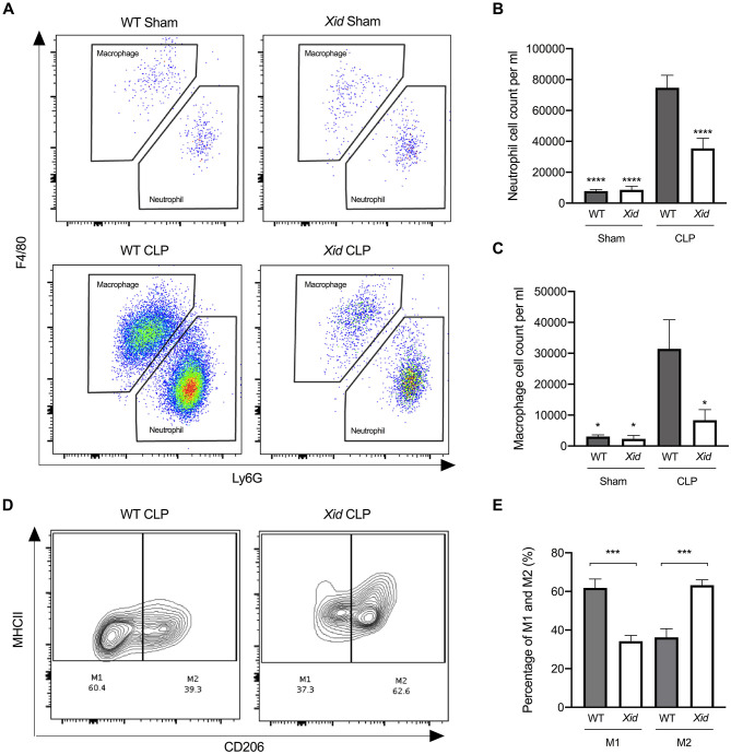
Figure 3.
Xid mice have fewer infiltrating immune cells in the peritoneum and enhanced polarization to M2 macrophages. Mice underwent sham or CLP surgery, 24 h later peritoneal lavage fluid was analyzed. (A) Scattergrams illustrating macrophage (identified as F4/80+Ly6G−) and neutrophils (identified as F4/80−Ly6G+). (B) Peritoneal neutrophil (F4/80−Ly6G+) cell count per ml. (C) Peritoneal macrophage (F4/80+Ly6G−) cell count per ml. (D) Contour plot illustrating percentage of M1 and M2 macrophages in WT and Xid mice, M1 identified as MHCII+CD206− and M2 identified as MHCII+CD206+. (E) Percentage of M1 and M2 macrophages in WT mice and Xid mice (%). The following groups were studied WT sham (n = 5), Xid sham (n = 5), WT-CLP (n = 10), and Xid-CLP (n = 10). Data are expressed as mean ± SEM and analyzed by one-way ANOVA with a Bonferroni post hoc-test. *P < 0.05, ***P < 0.001, and ****P < 0.0001 vs. WT-CLP.

