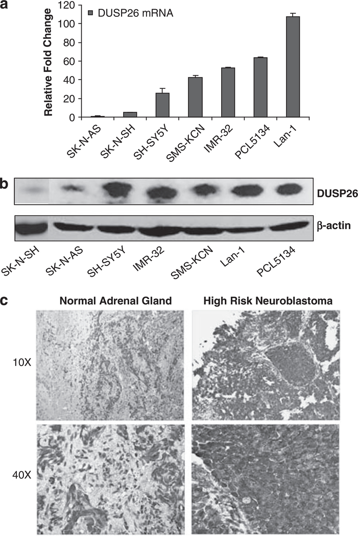
Figure 1.
DUSP26 expression in neuroblastoma cell lines and primary tumor specimens. (a) DUSP26 mRNA level in human neuroblastoma cell lines determined by quantitative reverse transcriptase (RT)–PCR. (b) DUSP26 protein level in human neuroblastoma cell lines determined by immunoblotting analysis. (c) Immunohistochemistry analysis of DUSP26 expression in primary neuroblastoma tumor specimens. Neuroblastoma paraffin-embedded tissues were stained with anti-DUSP26 antibodies and developed with DAB substrate and hematoxylin counterstain. Slides were viewed at × 10 and × 40 magnification.

