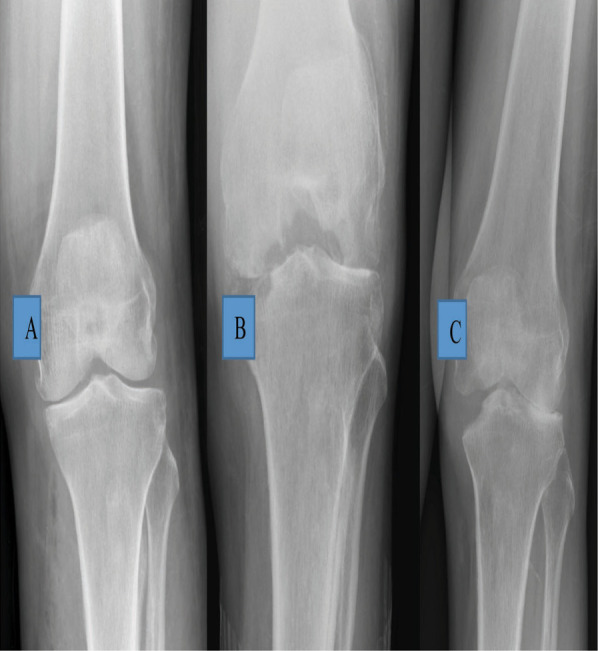
Figure 1-A., Figure 1-B., Figure 1-C. Radiograph (taken elsewhere) - AP view of the left knee just before the last intra-articular injection was given, showing that the medial joint space is narrowed, but the femoral and tibial condyles are not destroyed, Radiographs AP and lateral views of the same knee taken a month later, on admission, show marked destruction of femoral and tibial condyles. Such rapid destruction of bone is commonly seen due to pyogenic infection. Posterior subluxation of tibia seen in Lateral view, AP radiographs of the both knees 9 months post-arthrotomy and antibiotic therapy, shows some reformation of the medial femoral and tibial condyles.

