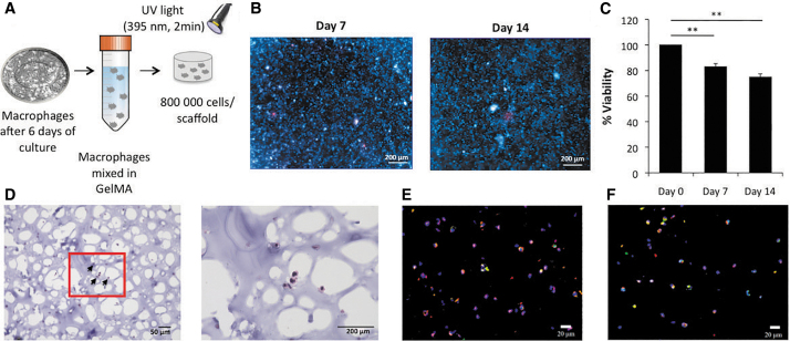
FIG. 2.
Macrophages in 3D culture. (A) Procedure for 3D culture of macrophages in GelMA. Monocytes were differentiated into macrophages for 6 days, trypsinized and pelleted. The macrophages were resuspended in GelMA solution and photocrosslinked to fabricate the 3D scaffolds. (B) Live (blue)/Dead (red) staining of constructs at day 7 and 14 (scale bar = 200 μm). (C) Cell viability quantification by PicoGreen DNA assay at days 7 and 14 (normalized to day 0 data). Each value represents the mean ± SD. **p < 0.01, compared with day 0. (D) Hematoxylin and Eosin staining at day 14 (scale bar = 50 μm [left] or 200 μm [right]). Arrows point to macrophages in the scaffold. (E) Immunofluorescence staining of CCR5 (red), CD71 (green), and DAPI nuclear staining (blue). (F) Immunostaining of CCR5 (red), CD68 (green), and DAPI nuclear staining (blue) at day 14 (scale bar = 20 μm). Each value represents the mean ± SD. **p < 0.01 compared with day 0. 3D, three-dimensional; SD, standard deviation; GelMA, methacrylated gelatin; DAPI, 4′,6-diamidino-2-phenylindole. Color images are available online.

