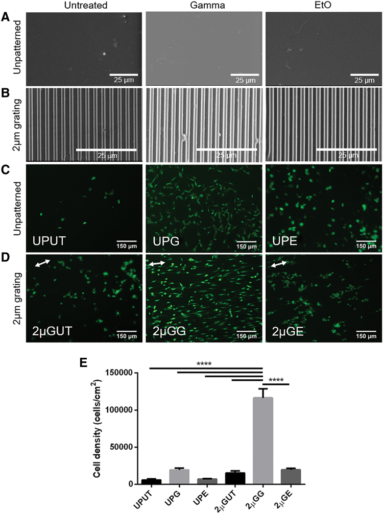
FIG. 6.
The effect of sterilization on cell adhesion and proliferation to unpatterned and patterned PVA films. SEM was performed on (A) unpatterned and (B) 2 μm grating PVA films before and after sterilization treatment. There was no noticeable difference in the texture of unpatterned surfaces of treated and untreated hydrogels. Gratings were also not affected by either γ or EtO treatment. Cells were seeded on these substrates and stained with live/dead assay after being cultured for 13 days; live cells were stained in green and dead cells were stained in red (no dead cells were found). (C) Unpatterned film: untreated control (UPUT), γ irradiated (UPG), and EtO treated (UPE). (D) Two micrometers grating film: untreated control (2 μGUT), γ irradiated (2 μGG), and EtO treated (2 μGE). White arrows indicate the parallel orientation of the microgratings. Scale bars represent 25 μm for SEM images and 150 μm for fluorescence images. (E) The effect of sterilization on the proliferation of cells as measured with CyQUANT assay. Quantification was performed after 13 days of culture. All data are presented as the average of n = 5 samples ± SE except for UPG and 2 μGUT where n = 4. ****Represents statistical significance with p ≤ 0.0001. SEM, scanning electron microscopy. Color images are available online.

