FIG. 8.
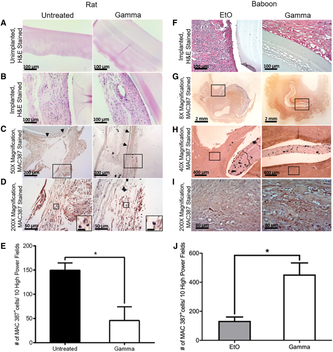
Histology of untreated versus γ-irradiated PVA films and EtO- versus γ-treated PVA grafts after being implanted subcutaneously on the dorsal side of a rat for 21 days and at the abdominal aorta iliac bifurcation of baboons for 28 days, respectively. H&E staining of (A) unimplanted film, (B) implanted film in rat, and (F) implanted graft in baboon. H&E and immunostaining with MAC387 antibody of (C) rat tissue surrounding the film implant, 50 × magnification, with area chosen for higher magnification bounded with rectangle and arrows pointing to the graft/tissue interface, (D) 200 × magnification of (C), with inserts showing the zoomed in images of the stained macrophages. Scale bar of inserts: 10 μm. (E) The total number of rat MAC 387+ macrophages per 10 high-power fields (200 × ) in the surrounding tissues of untreated and γ-irradiated grafts. H&E and immunostaining with MAC387 antibody of baboon tissue surrounding the graft implant with (G) 8 × , (H) 40 × , and (I) 200 × magnification. Area chosen for higher magnification was bounded with rectangles. (J) The total number of baboon MAC 387+ macrophages per 10 high-power fields (200 × ) in the surrounding tissues of EtO- and γ-irradiated grafts. Statistical significance was calculated with unpaired t-test (*p < 0.05; p = 0.0346). Brightness adjustment was performed using ToupView software. H&E, hematoxylin and eosin. Color images are available online.

