
Fig. 1.
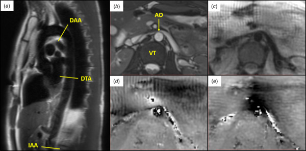
(a) Axial locations along the aorta analyzed by DENSE MRI. (IAA, DTA, and DAA) (b–e) Illustrative example of images acquired at peak systole in the infrarenal abdominal aorta of a healthy 23 year old female: (b) SSFP cine, (c) DENSE magnitude, (d) DENSE x-phase encoding, and (e) DENSE y-phase encoding. (AO—aorta and VT—vertebra)