Figure 1.

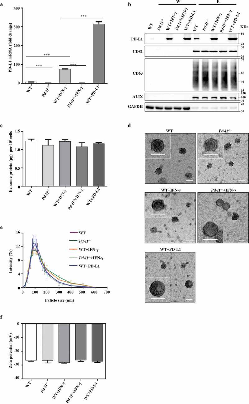
Characterization of exosomes purified from melanoma cells (a). qPCR of PD-L1 mRNA levels in SK-MEL-5 cells (WT), PD-L1 knockout SK-MEL-5 cells (Pd-l1−/-), IFN-γ treated cells (WT+IFN-γ, Pd-l1−/-+IFN-γ) and PD-L1 overexpressing cells (WT+PD-L1). n = 3. (b). Western blot for PD-L1, CD81, CD63, ALIX and GAPDH in the whole cell lysate (W) and purified exosomes (E) from WT, Pd-l1−/-, WT+IFN-γ, Pd-l1−/-+IFN-γ and WT+PD-L1. (c). The protein yield of exosomes from WT, Pd-l1−/-, WT+IFN-γ, Pd-l1−/- +IFN-γ and WT+PD-L1. n = 3. (d). TEM images of purified exosomes from WT, Pd-l1−/-, WT+IFN-γ, Pd-l1−/-+IFN-γ and WT+PD-L1. Scale bar: 50 nm. (e-f). The size distribution (e) and the Zeta potential (f) of exosomes from WT, Pd-l1−/-, WT+IFN-γ, Pd-l1−/-+IFN-γ and WT+PD-L1. n = 3. ***P < 0.001.
