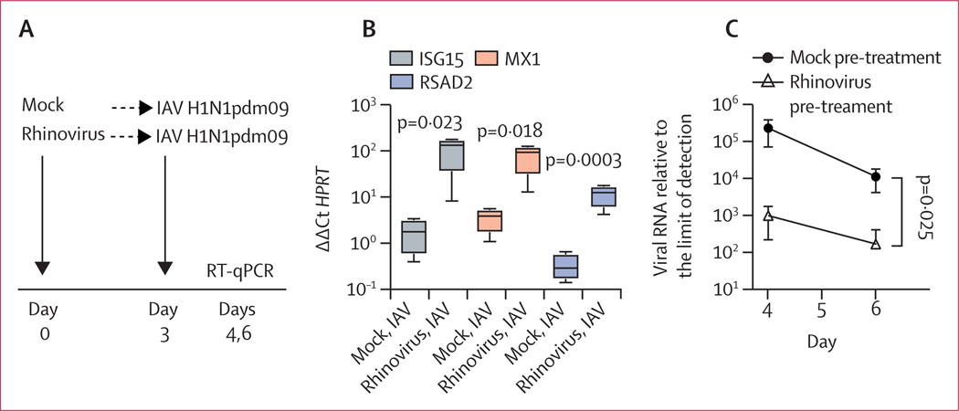
Figure 3: Effect of previous rhinovirus infection on epithelial gene expression and 2009 pandemic influenza A virus infection in differentiated airway epithelial cultures.
Data are mean (SD) of four replicates per condition. (A) Timing of sequential infections. (B) ISG mRNA expression on day 4, with or without previous rhinovirus infection. ISG expression amounts are graphed relative to the housekeeping gene HPRT. (C) Amount of IAV H1N1pdm09 viral RNA measured on days 4 and 6 with or without previous rhinovirus infection. The amount of viral RNA is expressed as fold change from the limit of detection. Significance of differences between mock pre-treated and rhinovirus pre-treated conditions were assessed by t-test (B) or two-way ANOVA (C). IAV=influenza A virus. ISG=interferon-stimulated gene. RT-qPCR=reverse-transcription quantitative PCR.

