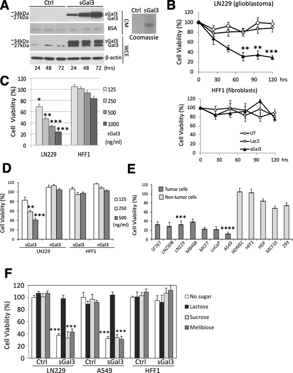
Figure 1: Secreted Gal-3 inhibits tumor cell viability in vitro.
A) Western blot showing levels of endogenous (Gal-3) and secreted (sGal-3) forms of Galectin-3. Left panel: cell extracts (WCE) and supernatants (CM) from 293 cells 48 hrs after transient transfection with an expression vector encoding LGALS3 cDNA fused to a classical secretion signal (pUMVC7) or control vector pCMV-LacZ (Ctrl.) were analyzed. BSA, Ponceau staining of bovine serum albumin (BSA). Right panel: Coomassie blue staining shows lactosyl-sepharose-purified sGal3 from CM.
B) Crystal violet cell viability assay showing tumor cell-specific toxicity of sGal-3. Human glioma cells (LN229, upper panel), and human fibroblasts (HFF-1, lower panel) were treated in triplicate for 30–120 hrs. with CM from 293 cells either untransfected (UT), or transfected with control (LacZ) or sGal3-expressing vectors. **p <0.01, ***p <0.001 (unpaired t-test).
C) Comparison between sGal-3 CM and rGal-3-mediated cytotoxicity in LN229 and HFF-1 cells after 48 hrs. treatment (upper panel). Quantification is presented as percent of sGal-3/vector control CM crystal violet staining from triplicates. *p <0.05, **p <0.01, ***p < 0.001 compared to control CM (unpaired t-test).
D) Dose-response of purified sGal-3 on LN229 and HFF-1 cell viability by crystal violet assay after 48 hrs. sGal-3 CM was purified through lactosyl-Sepharose column and quantified by ELISA. Quantification from triplicates as above. **p < 0.01 ***p < 0.001 (unpaired t-test). sGal-3 treatment of LN229 cells induced caspase-3 and PARP cleavage (lower panel).
E) Crystal violet assay demonstrating sGal-3 CM induced death at 48 hrs in genetically and biologically heterogeneous malignant human cancer cell lines (black) derived from brain (SF767, LN-Z308 and LN229), breast (MD468 and MCF7), lung (A549 and H1289) and prostate (LnCaP and PC3) tumors. In contrast, primary cultures of human endothelial cells (HDMEC) or fibroblasts (HDF and HFF-1) or normal breast epithelial cells (MCF10) or embryonic neuroepithelial 293 cells (grey) did not show significant decreases in cell viability. Cell viability is expressed as percentage sGal-3 over control CM. Three independent experiments were performed in triplicate (n=3). ***p < 0.001, ****p < 0.0001 compared to control CM (unpaired t-test).
F) Crystal violet assay showing neutralization of sGal-3 CM with lactose. sGal-3 CM was pre-treated for 1 hr with 20 mM (final concentration) of lactose, sucrose, and melibiose, then used to treat tumor (LN229, A549) and normal (HFF-1) cells for 48 hrs. Quantified as percent of sGal-3/control CM crystal violet staining from triplicates. ***p < 0.001 compared to control CM (unpaired t-test).

