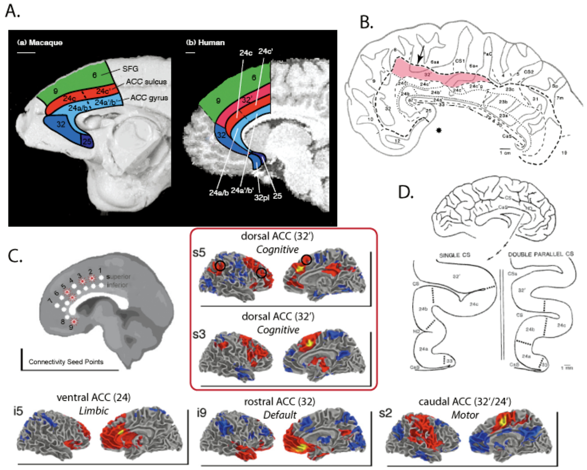
Figure 2 – Anatomy of Anterior Cingulate Cortex.

A) Macaque (left) and human (right) primate brains are illustrated with corresponding anatomical areas illustrated. Importantly, a dorsal-caudal extension of area 32, area 32′, is present only in the human brain. Figure adapted from [20]. B) A flatmap illustration of human anterior cingulate, illustrating the location and extent of area 32′. The arrow indicates that area 32′ often extends onto the dorsal medial wall surface. Figure adapted from [42]. C) Resting-state functional connectivity MRI maps of seed points along human ACC. Area 32′ extends from superior point 3 (s3) to s6, and shows connectivity with cognitive control regions posterior parietal cortex, DLPFC, and possibly nearby medial frontal regions. This region is separated connectively from caudal ACC in posterior area 32′/24′ (which appears to be a motor area), ventral ACC in area 24 (which appears to be a limbic area), and rostral ACC in area 32 (which is part of the “default state” network; [46]). Figure adapted from [45]. D) The anatomy of ACC is extremely variable between individuals. 30–50% of humans have a double cingulate in at least one hemisphere [24, 36]. Figure from [42].
