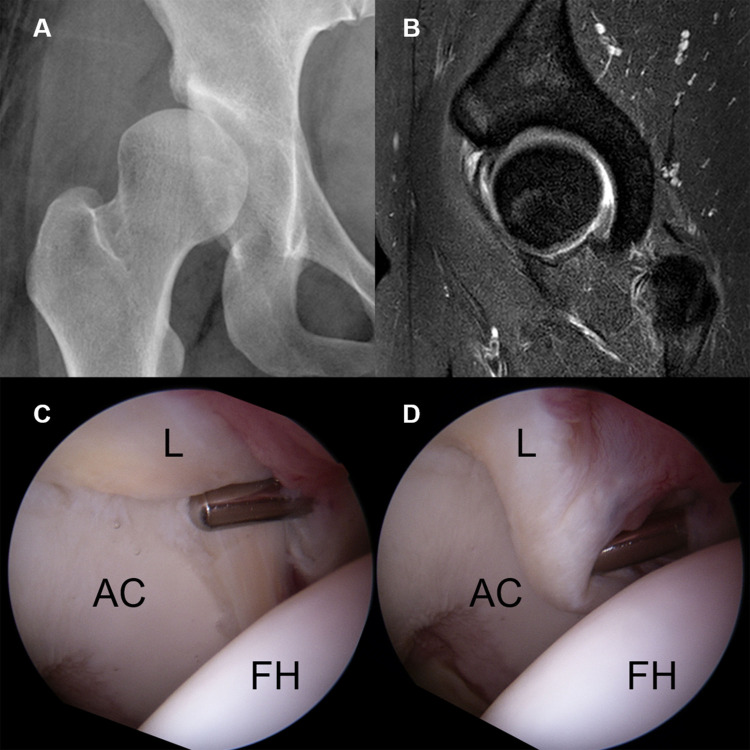
Fig 4. A grade 3 labral injury in a 26-year-old woman.
(A) Simple X-ray showing a definite dysplastic acetabular structure. (B) Sagittal hip MRI showing increased intrasubstance signal intensity and a contrast material-filled paralabral cyst at the chondrolabral junction. (C, D) Arthroscopy showing an unstable labrum between the chondrolabral junction (C) and the capsulolabral recess (D), without labral displacement. (L, labrum; FH, femoral head; AC, acetabular cartilage).

