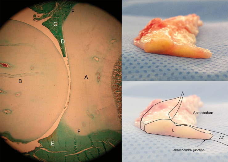
Fig 6.
Photomicrograph of a fetal hip at term (left, reprinted with permission from M. Cashin et al.: Embryology of the acetabular labral-chondral complex. J Bone Joint Surg [Br] 2008;90-B:1019–24. Copyright 2008 Springer.) (A, acetabulum; B, femoral head; C, anterior labrum; D, intra-articular projection of the anterior labrum; E, posterior labrum; F, acetabular labral transition zone). Gross specimen of a labrum (right top), resected during total hip arthroplasty from a patient with hip dysplasia, and a schematic drawing of the acetabulum including it (L, labrum; AC, acetabular cartilage).

