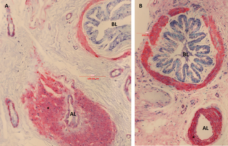
Fig 8.
Muscular pulmonary artery and annexed bronchus of an asthmatic horse in remission (A) and of a control horse (B) in study 2 (immunostained sections, 40X, α-smooth muscle actin). The bronchus lumen (BL) and artery lumen (AL) are identified. In these micrographs, significant increase in vascular smooth muscle (*) of the muscular pulmonary artery is visually detectable in the asthmatic horse (A), compared to a control (B).

