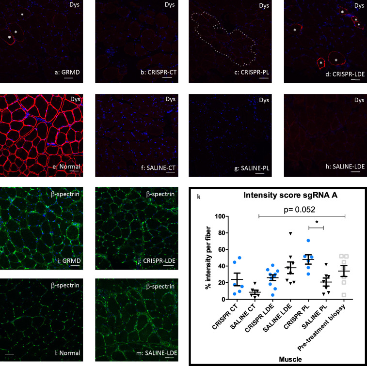
Fig 6. Dystrophin expression was minimally restored in HDR-CRISPR-Tx muscle from sgRNA A.
Dystrophin co-stained with C and N-terminus antibodies with Alexa 647 (red), β -spectrin membrane control (green) and DAPI denotes the nuclei (blue). Asterisks denote cells with a value of 2 in intensity score for dystrophin signal in the GRMD non-Tx and Tx samples. Dotted line denotes cells with a value of 1 in intensity score. Scale bar = 50μm. (a) Pre-treatment biopsy sample for Miercoles. (b) HDR-CRISPR injected cranial tibial (CT) Miercoles. (c) HDR-CRISPR injected peroneus longus (PL) Miercoles. (d) HDR-CRISPR injected long digital extensor (LDE) Miercoles. (e) Normal dog muscle. (f) SALINE injected CT Miercoles. (g) SALINE injected PL Miercoles. (h) SALINE injected LDE Miercoles. (i) Pre-treatment biopsy sample for Miercoles. (j) HDR-CRISPR injected LDE Miercoles. (k) Dystrophin intensity quantification for Miercoles and Friendly via One-way ANOVA multiple comparisons test, blue circle indicates CRISPR-Tx limb, black triangle indicates SALINE-Tx limb, gray square indicates pre-treatment biopsied sample; *p<0.05. (l) Normal dog muscle (m) SALINE injected LDE Miercoles. Dys = dystrophin; p = p. value.

