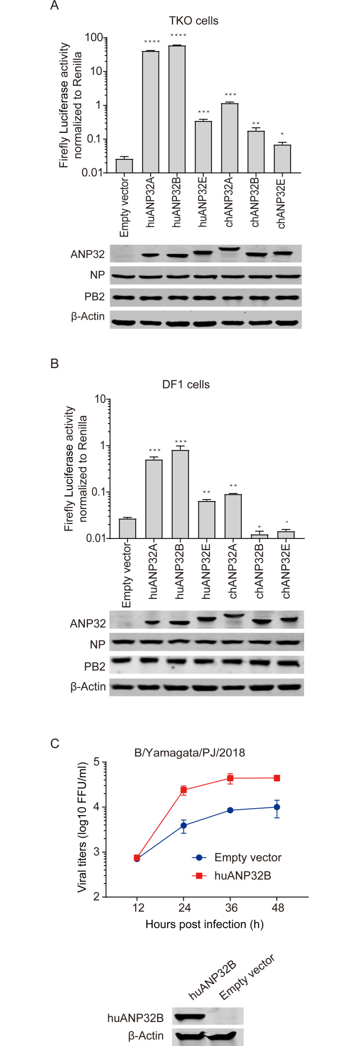
Fig 2. Species-specific support of influenza B viral polymerase activity by ANP32 proteins from different animals.
(A) TKO cells were co-transfected with B/Yamagata/1/73 polymerase, minigenome reporter, Renilla expression control and 10 ng of one of the following: huANP32A, huANP32B, huANP32E, chANP32A, chANP32B, chANP32E or 10 ng empty vector. Luciferase activity was assayed at 24 h after transfection. The expression of ANP32 proteins and polymerase was assessed using western blotting. (B) DF1 cells were co-transfected with B/Yamagata/1/73 polymerase, minigenome reporter with chicken polI promoter, Renilla expression control and 10 ng of one of the following: huANP32A-flag, huANP32B-flag, huANP32E-flag, chANP32A-flag, chANP32B-flag, chANP32E-flag or 10 ng empty vectors. Luciferase activity was assayed at 24 h after transfection. The protein expression was determined by western blotting using different antibodies: anti-flag antibody for ANP32 proteins, and specific antibodies to polymerase and β-actin. The data indicate the firefly activity normalized to Renilla, Statistical differences between cells are labeled according to a one-way ANOVA followed by a Dunnett’s test (NS = not significant, **P < 0.01, ***P < 0.001, ****P < 0.0001). Error bars represent the SD of the replicates within one representative experiment. (C) DF1 cells were transfected with 1 μg huANP32B-flag or empty vector in 6 well plate. Twenty-four hours post transfection DF1 cells were infected with B/Yamagata/PJ/2018 virus at a MOI of 0.1 and cultured at 33°C or 37°C. The supernatants were sampled at 12, 24, 36, 48 h post infection and the viral titers were determined using Fluorescence Focus Units (FFU) assay on MDCK cells. The expression of huANP32B was assessed by western blotting using anti-flag antibody. The result is shown as average of n = 3 ± SD.

