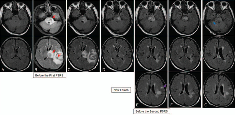
Figure 2.

Cranial magnetic resonance imaging (MRI) scans revealed the sizes of brain metastases (BM) lesions and peritumoral brain edema (PTBE). (A) No metastatic lesion observed prior to surgery in March 2018; (B) 3 BM (red arrow indicates left temporal-parietal lobe, left thalamus lesions, and brain stem lesion), and large areas of PTBE observed in June 2018; (C) the BM lesions were reduced, and the area of the PTBE shrank progressively after radiation therapy; (D) all BM stayed reduced, and PTBE had disappeared in October 2018; (E) cranial MRI scans showed a new BM lesion in the left frontal lobe (purple arrow), and there were no obvious changes in the 3 BM in January 2019; (F) all BM lesions appeared further reduced in March 2019; (G) cranial MRI scans showed a new BM (blue arrow) in the cerebellum in October 2019.
