
Fig. 2.
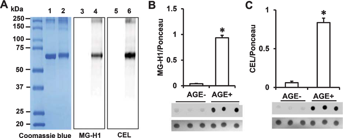
Determination of AGE adducts in the AGE additive and AGE diet. A) Protein (2 μg) from BSA or MG-BSA samples following 12 days of incubation were subjected to SDS-PAGE. Protein bands were stained by Coomassie Blue (A, Lane 1 & 2). Glycation was detected by immunoblotting with antibody to MG-H1 (A, Lane 3 & 4) or CEL (A, Lane 5 & 6). Lane 1, 3, 5, BSA; lane 2, 4, 6, MG-BSA. Quantification of immunodot intensity of MG-H1 (B), and CEL (C) normalized to Ponceau staining intensity (as a protein loading control) in protein extracted from the samples of AGE− and AGE+ diet. Data are presented as mean ± SEM (n = 3), *p < 0.01 versus AGE− diet.