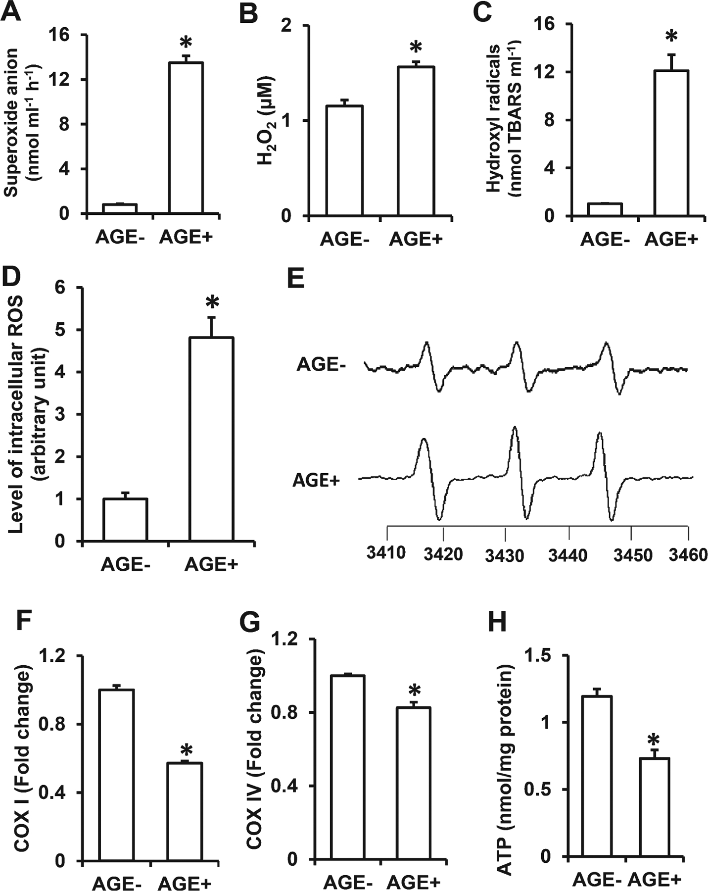
Fig. 5.

Effect of AGE diet on ROS generation and mitochondrial function. Quantification of superoxide anion (A), hydrogen peroxide (H2O2) (B), hydroxyl radicals (C) in serum of AGE− and AGE+ fed mice. Representative in vitro EPR spectra measured in cortical brain homogenates (D, E) are shown. The peak height in the spectrum indicates levels of ROS. Quantification of EPR spectra in the indicated AGE− & AGE+ fed mice (D). *p < 0.01 compared to other groups of mice. Data are expressed as fold-increase relative to AGE− fed mice. N = 5 mice per group. Activities of complexes I (F) and complex IV (G) and ATP levels (H) were determined in the cortex of AGE− and AGE+ mice. Data presented as mean ± SEM (n = 5), *p < 0.01 versus AGE− control group.
