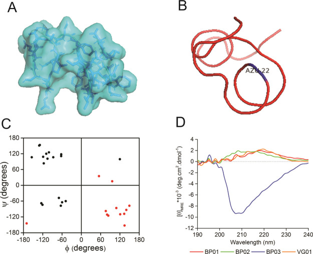
Figure 2.
Representation of (A) surface model, (B) C-alpha traces, and (C) dihedral angles of the protein foldamer BP02, where d-amino acids are shown in red circles and l-amino acids are shown in black circles. The representation in all four quadrants of the Ramachandran map confirms the heterochiral nature of the designed protein. (D) CD spectra of the designed proteins indicating the disordered conformation of the designed proteins.

