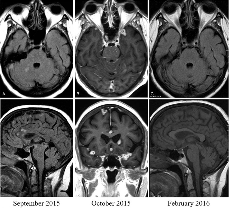
Figure 1.

Magnetic resonance imaging of case 1. Axial and sagittal fluid-attenuated inversion recovery images without gadolinium (A) and axial and coronal T1-weighted images with gadolinium (B) performed 5 (September 2015) and 8 weeks (October 2015) respectively after starting tuberculosis treatment, showing multiple supra-and infratentorial lesions, distributed in the brain parenchyma and subarachnoid space. Axial and sagittal scans without gadolinium (C), taken after 6 weeks after receiving a three-dose course of infliximab, showing complete resolution of the tuberculomas.
