
FIGURE 2.
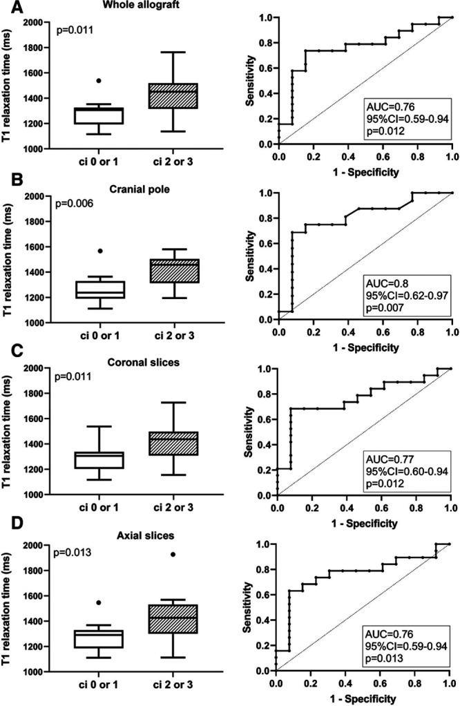
Results of T1 mapping in relation to groups defined according to Banff ci scoring. Cortical T1 relaxation times are shown for the whole transplant (A) and the cranial pole (B), as well as coronal (C) and axial (D) sections of the renal allograft. Box plots indicate the median, interquartile range, and the minimum and maximum of T1 values. Outliers are indicated as circles. The Mann–Whitney U test was used for group comparisons. The prediction of ci scores >1 by T1 mapping results is presented by receiver operating characteristic (ROC) curves and the corresponding area under the curve (AUC) including 95% confidence interval (CI) and P values.