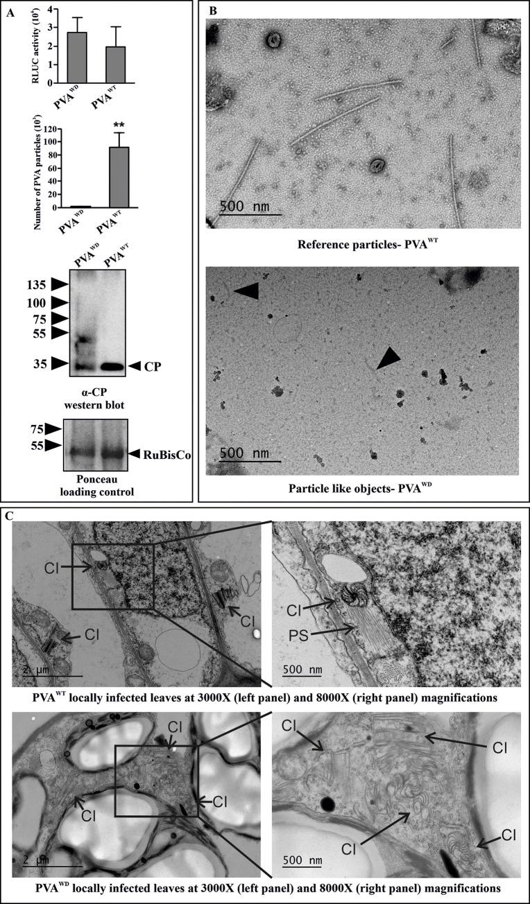
Fig 8. PVAWD is defective in particle formation.
(A) PVA particle accumulation in PVAWD and PVAWT infected local leaves at 3 dpi, as measured by immunocapture qRT-PCR. N. benthamiana plants were agroinfiltrated with PVAWD (OD600 = 1) and PVAWT (OD600 = 0.1) to ensure comparable RLUC expression level between PVAWD and PVAWT (upper panel). The corresponding particle numbers and CP accumulation visualized by western blotting with anti-CP antibodies are presented in the middle and lower panels, respectively. (B) Electron microscopy images from the same samples as in A) showing normal virus particles formed by PVAWT. No particles were found in PVAWD, only few deformed particle-like structures were observed (marked by an arrow in the lower electron micrograph). The number of plants per experiment was 3. Statistical significance was assessed using student’s t-test (*P < 0.05). (C) Both PVAWT and PVAWD infections were allowed to spread in the local leaves until 9 dpi, and EM images of the infected tissues were taken.Pinwheel inclusions (marked as ‘CI’) were observed. However, the pinwheels from PVAWT were associated with stacks of particles (marked as ‘PS’) in vicinity while those from PVAWD were devoid of them.

