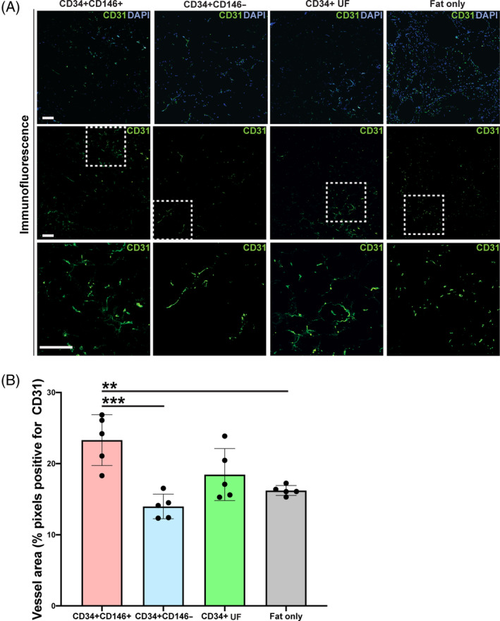
FIGURE 6.

Vascularization of explanted fat grafts 8 weeks postgrafting. A, Immunofluorescence staining for endothelium using CD31 (green) in fat grafts enriched with CD34+CD146+ (far left), CD34+CD146− (middle left), CD34+ UF (middle right) ASCs, or not enriched alone (“fat only,” far right) shown at low magnification with DAPI (blue) (top row), with CD31 alone (middle row), and of the selected ROI (white dotted box) at high magnification (bottom row). Scale bars = 100 μm. B, Fat graft vascularization was quantified as ROI area occupied by CD31‐positive pixels. Fat enriched with CD34+CD146+ ASCs had greatest vascularization, indicated by increased CD31 immunofluorescence staining compared to fat enriched with CD34+CD146− or CD34+ UF ASCs, and grafts not enriched with ASCs and fat alone; n = 5 per group (*P < .05, **P < .01). ASC, adipose‐derived stromal cell; DAPI, 4′,6‐diamidino‐2‐phenylindole; ROI, region of interest; UF, unfractionated
