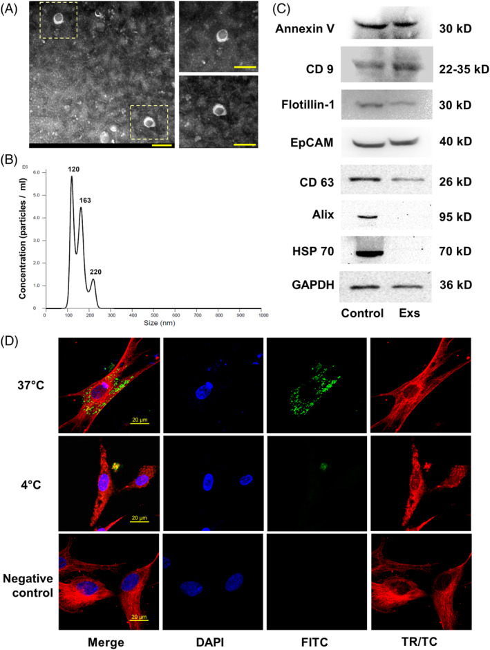
FIGURE 2.

Characterization of exosomes derived from P2X7R gene‐modified stem cells. A, Representative TEM image of exosomes (scale bar = 200 nm). B, Size distribution profile of exosomes as determined by nanoparticle tracking analysis. C, Western blot analysis of cell lysates (control) and exosomes (Exs) showing the presence of the marker proteins Annexin V, CD 9, Flotillin‐1, EpCAM, and CD63 in both cell lysates and exosomes, while exosomes were negative for Alix and HSP70. D, Representative confocal image of fluorescently labeled exosomes (green) endocytosed by PDLSCs at 37°C or 4°C counterstained with tubulin (red) (scale bar = 20 μm). PDLSC, periodontal ligament stem cell; TEM, transmission electron microscopy
